Chiropractic Biophysics (CBP) Lakewood Colorado
CBP® Goals of Care
CBP® Technique emphasizes optimal posture and spinal alignment as the primary goals of chiropractic care while simultaneously documenting improvements in pain and functional based outcomes (See Figure 1). The uniqueness of CBP® treatment is in structural rehabilitation of the spine and posture. In general the goals of CBP® Care are:
- Normal Front & Side View Posture
- Center of mass of head, rib cage & pelvis vertically aligned in Front and Side views.
- Normal Spinal Alignment
- Front view: vertical alignment
- Side View: Harrison Ideal or Average Spinal Model
- Normal function
- Improved Range of Motion and quality of movement,
- Improved muscle strength,
- Improved Health & Symptom Improvements
- Neck disability index
- Oswestry low back index,
- SF 36 or Health Status Questionnaire
Ideal Postural Alignment:
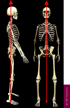
Figure 1. Ideal postural alignment is depicted in both the frontal and side views. In each view, the center of mass of the skull, thorax, and pelvis are in a vertical line with respect to gravity. In the frontal view, the spinal column is vertically aligned-a straight column- with respect to gravity. In the side view, the spine has three primary curvatures which will be described below:
1. Neck Curve – Cervical Lordosis,
2. Ribcage Curve – Thoracic Kyphosis,
3. Low back Curve – Lumbar Lordosis
Ideal Spinal Alignment: Harrison Full Spine Model
As in all fields of study dealing with the human body, i.e. physiology, hematology, anatomy, etc., there exist normal values for alignment of the spine. The Harrison Spinal Model is an evidenced based model for side view spinal alignment. It is the geometric path of the posterior longitudinal ligament or the backs of the vertebra from the 1st neck vertebra to the bottom of the lower back or top of the sacrum. See Figures 2-6 below detailing the Harrison Spinal Model.
CBP® researchers have extensively published ideal and average models for the human spinal curvatures as viewed from the side. This research has lead to the finding of the ‘Harrison Spinal Model’. This model details both Ideal and Average geometric shapes for the curves of the spine from the side. Additionally, ideal and average ranges for the spinal segmental angles for each of the spinal regions have been identified. The neck or cervical spine should have a geometric shape that approximates a ‘piece of a circle’. The ribcage or thoracic spine should have a geometric shape that approximates an oval-elliptical shape. And the low back or lumbar spine should have a geometric shape that approximates an oval-elliptical shape.26-31
These are “evidence based” models. In fact, the CBP® neck-cervical circular model27 and the low back- lumbar elliptical model29 have both been found to have discriminative validity between pain and non pain subjects. In other words, the Harrison Spinal Model has been found to be able identify pain subjects versus non-pain subjects by what their spinal x-ray shapes are.
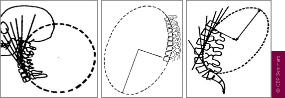
Figure 3. These three figures demonstrate the concept that each spinal region has a normal geometry or shape of the spinal curves. On the readers left is the Neck or Cervical spine-Here the shape in the neck curve should approximate a piece of a circle. In the Center is the Ribcage or Thoracic spineHere the shape in the ribcage should approximate a piece of an oval or ellipse. On the Right is the Low back or Lumbar spine. Here the shape in the low back should approximate a piece of an oval or ellipse.
F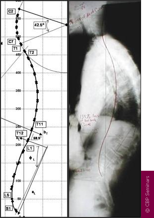 igure 4. The Harrison Full Spine Model. On the readers left is the exact geometric model of the side view of the spinal curves as identified by Harrison and colleagues. This model can be used to determine what is wrong-abnormal with a given patients side view of the spine. For example, a full spine x-ray on the right is shown. The red-curved line represents the Harrison spinal model and this shows where the patients spinal vertebra should be lined up. It is apparent that this patient has altered spinal alignment as they do not fit even close to the Harrison Idealized Spinal Model.
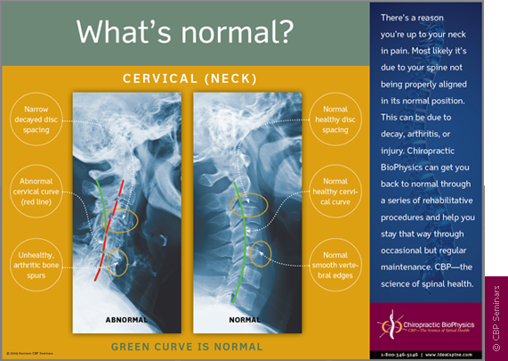
Figure 5. The Harrison Spinal Model in the Neck-Cervical region. The Harrison spinal model is depicted as the GREEN curved line in this figure. On the Right is a normal curved patient x-ray. On the Left is an abnormal curved patient x-ray; where the patients abnormal shape is shown by the Red dashed line. The Harrison Spinal Model in the neck has been shown to reasonably predict which person will have neck pain compared to normal subjects.
Figure 6. The Harrison Spinal Model in the Low back or Lumbar region. The Harrison spinal model is depicted as the RED curved line in this figure. On the Right is a normal curved patient x-ray. On the Left is an abnormal curved patient x-ray; where the patients abnormal shape is shown by the faint dashed line. The Harrison Spinal Model in the low back has been shown to reasonably predict which person will have low back pain compared to normal subjects.
X-Ray Analysis and Utilization
To establish optimal and average sagittal models, x-ray analysis and line drawing procedures are utilized. CBP® protocols require that the doctor must measure the displacements on spinal radiographs (segmental Subluxation). Both lateral-side view and anterior to posterior (AP) or frontal view CBP® x-ray line drawing procedures have been studied and found to be reliable.32-36 Furthermore, CBP® utilizes standardized x-ray positioning procedures that have been studied and found to be reliable.36
As with measures of pain intensity, range of motion, and quality of life, periodic assessment of spinal structural alignment is important to evaluate progress and determine when maximum patient improvement has been reached. In CBP® Technique, the use of initial and follow-up spinal x-rays or radiographs is deemed necessary; however, some in chiropractic have condemned the use of follow-radiographs to collect alignment data.37-39 Importantly, there is data to show that the use of medical/chiropractic x-rays constitutes a very minor health risk and in fact has been shown to be of benefit (decreased sickness and cancer mortality rates) in some studies.40-42
In reality, the only way to see what an individual patients spine alignment looks likes, is to obtain spinal imaging such as Radiography or X-ray. No-one would not take their car to the mechanic and say: Somethings wrong with my engine but dont look under the hood–Would you? Then why would anyone want a Chiropractor to adjust-treat their spine without having an x-ray to see what the persons spine looked like? Would You?
CBP® Postural Analysis
Previously, engineering concepts were used to describe all spinal-vertebral segmental movements as rotations and translations in 3-dimensions.10 However, Dr. Don Harrison was the first to describe abnormal postures of the head, rib cage, and pelvis in this manner.1-3,9 Figures 7 and 8 depict the twelve simple motions in six-degrees of freedom as rotations and translations of the human head, ribcage, and pelvis:
• Rotation is a turning, twisting, or tilting movement and is an angular movement
• Translation is a straight line movement (up, down, left, right, forward, backward)
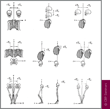
Figure 7. The possible translation postures (Tx, Ty, Tz) of the head, rib cage, and pelvis are depicted in 3-dimensions. In 1980, Dr. Don Harrison termed these pairs on any one axis as Mirror Images®. Whichever Abnormal postures were found to exist in the patient, these postures would be placed into their Mirror Image® before a force was applied with an adjusting instrument, drop table, exercise and/or traction.
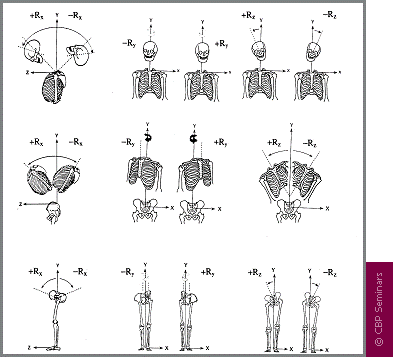
Figure 8. The possible postural rotations (Rx, Ry, Rz) of the head rib cage and pelvis are depicted in 3-dimensions. In 1980, Dr. Don Harrison termed these pairs on any one axis as Mirror Images®. Whichever postures were found to exist in the patient, these postures would be placed into their Mirror Image® before a force was applied with an adjusting instrument, drop table, exercise and/or traction.
The postural and spinal displacements are the determining factors for deriving a patients individualized program of care. Prior to performing CBP® Mirror Image® postural set-ups, the patients initial presenting abnormal posture(s) must be exactly determined.
Medical Disclaimer
The content on this website, renew-chiropractic.com, is not intended to be a substitute for professional medical advice, diagnosis, or treatment. Always seek the advice of your physician or other qualified health provider with any questions you may have regarding a medical condition. Never disregard professional medical advice or delay in seeking it because of something you have read on this website.
The information included on this site is for educational purposes only. It is not intended nor implied to be a substitute for professional medical advice. The reader should always consult his or her healthcare provider to determine the appropriateness of the information for their own situation or if they have any questions regarding a medical condition or treatment plan. Reading the information on this website does not create a physician-patient relationship. Renew Chiropractic assumes no responsibility or liability for how readers of this site choose to use the information contained within.
If you think you may have a medical emergency, call your doctor, go to the emergency department, or call 911 immediately.
These articles and recommendations are not tailored for the health of one particular individual and should not be considered professional consultation. Individuals who believe they are experiencing symptoms of an illness should always consult a doctor before beginning any form of treatment.
The opinions expressed on the Site and by Renew Chiropractic, P.C. are published for educational and informational purposes only, and are not intended as a diagnosis, treatment, cure or as a substitute for professional medical advice, diagnosis and treatment. Please consult a local physician, medical doctor or other health care professional for your specific health care and/or medical needs or concerns.
